Home
/ Anatomy Of Back Of Neck / Bony Anatomy Of The Neck Ent Clinic Sydney - The anterior jugular vein (v.
Anatomy Of Back Of Neck / Bony Anatomy Of The Neck Ent Clinic Sydney - The anterior jugular vein (v.
Anatomy Of Back Of Neck / Bony Anatomy Of The Neck Ent Clinic Sydney - The anterior jugular vein (v.. If you'd like to support us and get something great in return, check out our osce checklist booklet containing over 120 osce checklists head & neck anatomy. Anatomy of the hand overview. Our neck is where we find the seven cervical vertebrae, with c7 (the seventh cervical vertebra) meeting t1 (the first thoracic vertebra) at the base of the neck. 12 photos of the anatomy of the back of the neck. Magnetic resonance imaging of the head and neck.
A fractured neck of femur (nof) is a common orthopaedic presentation. This entry was posted in anatomy by admin. In the neck, the platysma when contracted throws the skin into oblique ridges parallel with the fasciculi of the muscle. A collection of anatomy notes covering the key anatomy concepts that medical students need to learn. The pll starts at c2 and goes down the back of the vertebral bodies and intervertebral discs.
Upper Cervical Spine Disorders Anatomy Of The Head And Upper Neck from www.spineuniverse.com From a topographical standpoint, there are six major muscle groups in the neck. This entry was posted in anatomy by admin. Dummies has always stood for taking on complex concepts and making them easy to understand. Jugularis they unite with small veins from the deep muscles at the upper part of the back of the neck, and form a vessel which enters the foramen in the transverse. The physicians originally studying human anatomy thought the skull looked like an helmet. Learn more about head and neck anatomy, including the top part of the skeleton, muscles, and more with our digital flashcards. From the sides and the back of the neck, the splenius capitis inserts onto the head region, and the splenius cervicis extends onto the cervical region. The anterior jugular vein (v.
Clinically, surface anatomy is used to split the neck into anterior and posterior triangles which provide clues as to the location of specific structures.
Head and neck anatomy is important when considering pathology affecting the same area. Despite being a relatively small region, it contains a range of important anatomical features. Traditionally the anatomy of the infrahyoid neck has been subdivided into a group of surgical triangles whose borders are readily palpable bones and. We've largely focused on the physical aspect of our spinal anatomy in this series. 3d human anatomy torso back muscles stock illustration 470591129. Neck muscles help support the cervical spine and contribute to movements of the head, neck, upper back, and posterior longitudinal ligament (pll). Anatomical principles underlying cranial nerve lesions; « back show on map ». From the sides and the back of the neck, the splenius capitis inserts onto the head region, and the splenius cervicis extends onto the cervical region. Whether it's to pass that big test, qualify for that big promotion or even master that cooking technique; Demonstrate sound knowledge of the surface/living and radiological anatomy of the head, neck and. Over 65000 hip fractures each year are recorded occur in the uk alone and they are becoming in this article, we will look at the classification, anatomy, clinical and radiological features, and management of neck of femur fractures. A collection of anatomy notes covering the key anatomy concepts that medical students need to learn.
Despite being a relatively small region, it contains a range of important anatomical features. The anterior jugular vein (v. Anatomy of male back and neck pain in blue stock. The head rests on the top part of the vertebral column, with the skull joining at c1. From a topographical standpoint, there are six major muscle groups in the neck.
Human Neck And Back Anatomy Bild Kaufen 12063721 Science Photo Library from media02.stockfood.com A collection of anatomy notes covering the key anatomy concepts that medical students need to learn. The neck muscles, including the sternocleidomastoid and the trapezius, are responsible for the gross motor movement in the muscular system of the head and neck. Dummies has always stood for taking on complex concepts and making them easy to understand. 12 photos of the anatomy of the back of the neck. 3d human anatomy torso back muscles stock illustration 470591129. Spine anatomy back neck pain tyler tx. Magnetic resonance imaging of the head and neck. The physicians originally studying human anatomy thought the skull looked like an helmet.
Neck, in land vertebrates, the portion of the body joining the head to the shoulders and chest.
Learn everything about the neck anatomy with this topic page. Whether it's to pass that big test, qualify for that big promotion or even master that cooking technique; Spine anatomy back neck pain tyler tx. Dummies helps everyone be more knowledgeable and confident in applying what they know. The neck muscles, including the sternocleidomastoid and the trapezius, are responsible for the gross motor movement in the muscular system of the head and neck. The neck is the area between the skull base and the clavicles. If you'd like to support us and get something great in return, check out our osce checklist booklet containing over 120 osce checklists head & neck anatomy. Anatomy of the back top image row 2: The pll starts at c2 and goes down the back of the vertebral bodies and intervertebral discs. 12 photos of the anatomy of the back of the neck. Learn about the various causes of back pain, including different kinds of arthritis. The structure is, of course, an important part of the conversation. Anatomy of the nervous system.
Despite being a relatively small region, it contains a range of important anatomical features. The cervical spine supports the weight and movement of your head and protects the nerves exiting your brain. Head and neck anatomy is important when considering pathology affecting the same area. Our neck is where we find the seven cervical vertebrae, with c7 (the seventh cervical vertebra) meeting t1 (the first thoracic vertebra) at the base of the neck. Your neck is like no other part of the vertebral spinal column and enables your head and neck a wide range of motion.
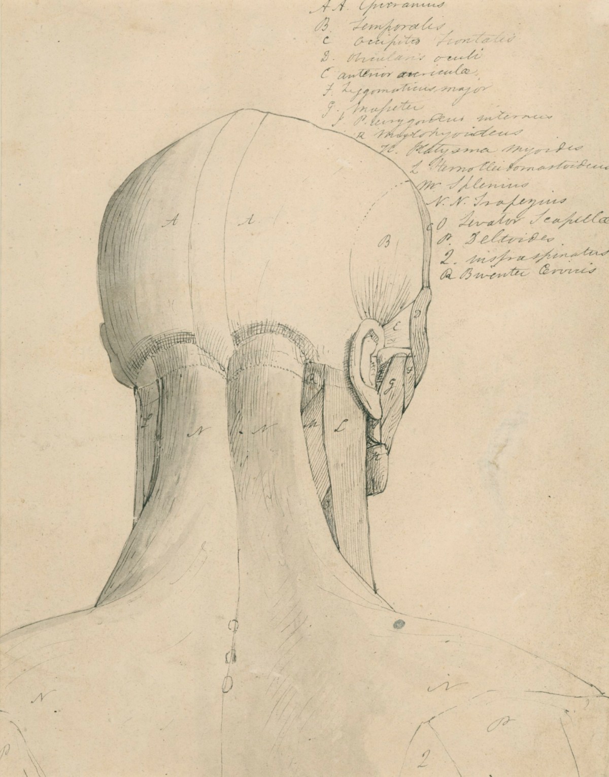
Anatomical Drawing Of The Back Of The Head And Neck Works Of Art Ra Collection Royal Academy Of Arts from d1inegp6v2yuxm.cloudfront.net Anatomy of the hand overview. 512 anatomical structures were dynamically labeled, and some structures have been redesigned or enhanced with a graphic tablet for better readability. Traditionally the anatomy of the infrahyoid neck has been subdivided into a group of surgical triangles whose borders are readily palpable bones and. Some important structures contained in or passing through the neck include the seven cervical vertebrae and enclosed spinal cord, the jugular veins and carotid arteries, part of the esophagus, the larynx. A fractured neck of femur (nof) is a common orthopaedic presentation. The cervical spine supports the weight and movement of your head and protects the nerves exiting your brain. From the sides and the back of the neck, the splenius capitis inserts onto the head region, and the splenius. The head rests on the top part of the vertebral column, with the skull joining at c1.
Anatomy of the back top image row 2:
The physicians originally studying human anatomy thought the skull looked like an helmet. The infrahyoid neck is the region of the neck extending from the hyoid bone to the thoracic inlet. The neck muscles, including the sternocleidomastoid and the trapezius, are responsible for the gross motor movement in the muscular system of the head and neck. If you'd like to support us and get something great in return, check out our osce checklist booklet containing over 120 osce checklists head & neck anatomy. Neck muscles help support the cervical spine and contribute to movements of the head, neck, upper back, and posterior longitudinal ligament (pll). The head rests on the top part of the vertebral column, with the skull joining at c1. Some important structures contained in or passing through the neck include the seven cervical vertebrae and enclosed spinal cord, the jugular veins and carotid arteries, part of the esophagus, the larynx. Want to learn more about it? 3d video tutorials and interactive modules on the anatomy of the back including anatomy of the musculature, vertebral column, joints and ligaments. Develop students understanding of the ways in which structure and function of muscle and joints. Demonstrate sound knowledge of the surface/living and radiological anatomy of the head, neck and. 12 photos of the anatomy of the back of the neck. « back show on map ».

